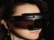
Compared with fluoroscopic guidance, VR reduces time to select GDA, splenic, right hepatic artery
THURSDAY, March 28, 2019 (HealthDay News) — Virtual reality (VR) can reduce the time for a catheter to reach target vessels, according to a study presented at the annual meeting of the Society of Interventional Radiology, held from March 23 to 28 in Austin, Texas.
Wayne Monsky, M.D., Ph.D., from the University of Washington in Seattle, and colleagues examined the feasibility of using a catheter with electromagnetic (EM) sensors projected onto a VR headset to see and steer the catheter through blood vessels. A three-dimensional (3D) hologram of the abdominal/pelvic vasculature was created from computed tomography angiography. A 3D printed model of the vasculature was held in anatomic orientation in a scaffold; a catheter and microcatheter with EM sensors were advanced in the 3D printed model. Using VR guidance, then using fluoroscopic guidance, the right hepatic, splenic, and gastroduodenal arteries (GDA) were selected six times each. The time taken to steer the catheter to the target vessels was compared.
The researchers found that when using VR display versus fluoroscopic guidance, the mean time to select the GDA, splenic, and right hepatic artery was 17.6, 18.6, and 22.6 seconds versus 70.3, 66.1, and 73.5 seconds, respectively. The mean time to select these vessels during clinical cases was 171.2, 92.3, and 188.4 seconds, respectively.
“Virtual reality will change how we look at a patient’s anatomy during an interventional radiology treatment,” Monsky said in a statement. “This technology will allow physicians to travel inside a patient’s body.”
Copyright © 2019 HealthDay. All rights reserved.


