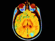
Findings seen in 23 children with congenital infection, presumably associated with Zika virus
FRIDAY, April 15, 2016 (HealthDay News) — Children with congenital infection, presumably associated with the Zika virus, have severe cerebral damage identified on imaging, according to a study published online April 13 in The BMJ.
Maria de Fatima Vasco Aragao, M.D., from the Mauricio de Nassau University in Brazil in Recife, and colleagues reported radiologic findings from computed tomography (CT) and magnetic resonance imaging (MRI) brain scans in 23 children with a diagnosis of congenital infection, presumably associated with the Zika virus.
The researchers found that six of the children tested positive for immunoglobulin (Ig)M antibodies to Zika virus in the cerebrospinal fluid. The other 17 children were not tested for IgM antibodies, but met the protocol criteria for congenital infection presumably associated with Zika virus. Fifteen of the children underwent CT, seven underwent CT and MRI, and one underwent MRI only. All 22 children who underwent CT had calcifications in the junction between cortical and subcortical white matter; and 21, 20, 19, and 11, respectively, had malformations of cortical development, decreased brain volume, ventriculomegaly, and hypoplasia of the cerebellum or brainstem. All eight children who underwent MRI had calcifications in the junction between cortical and subcortical white matter, malformations of cortical development (mainly in the frontal lobe), and ventriculomegaly.
“This study shows the largest and most detailed case series of neuroimaging findings in children with microcephaly and presumed Zika virus-related infection to date,” the authors write.
Full Text
Copyright © 2016 HealthDay. All rights reserved.


