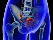
Parity tied to increased fiber length in more proximal coccygeus, iliococcygeus pelvic floor muscles
WEDNESDAY, Sept. 14, 2016 (HealthDay News) — Parity is associated with increased fiber length in the more proximal coccygeus and iliococcygeus pelvic floor muscles, according to a study published in the September issue of the American Journal of Obstetrics and Gynecology.
Marianna Alperin, M.D., from the University of California in San Diego, and colleagues examined the impact of vaginal deliveries and aging on human cadaveric pelvic floor muscle architecture. They obtained coccygeus, iliococcygeus, and pubovisceralis from younger and older donors, who were vaginally parous and vaginally nulliparous, all of whom had no history of pelvic floor disorders. Validated methods were used to assess architectural parameters.
The researchers found that the key impact of parity was increased fiber length in the more proximal coccygeus and iliococcygeus (P = 0.03 and 0.04, respectively). Across all pelvic floor muscles, aging changes manifested as decreased physiologic cross-sectional area (P < 0.05), which exceeded the age-linked decline in muscle mass. Younger vaginally parous pelvic floor muscles had lower physiologic cross-sectional area compared with younger vaginally nulliparous; the difference did not reach statistical significance. Collagen content of pelvic floor muscles was not altered by parity but increased with aging (P < 0.05).
“Increased fiber length in more proximal pelvic floor muscles likely represents an adaptive response to the chronically increased load placed on these muscles by the displaced apical structures, presumably as a consequence of vaginal delivery,” the authors write.
Copyright © 2016 HealthDay. All rights reserved.


