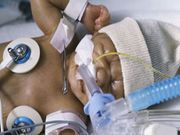
Brain scans shortly after birth may pinpoint potential developmental problems
THURSDAY, Jan. 19, 2017 (HealthDay News) — Magnetic resonance imaging (MRI) shortly after birth might help determine which premature babies have sustained a brain injury that will affect their development, according to a study published online Jan. 18 in Neurology.
Steven Miller, M.D., C.M., head of neurology at the Hospital for Sick Children in Toronto, and colleagues performed MRI scans on 216 infants born at an average 28 weeks of gestation. The investigators assessed both the volume and location of white matter injury. The research team then revisited 58 of the babies at 18 months of age, performing motor, cognitive, and language assessments to determine how their development had proceeded.
These assessments revealed that damage in the frontal lobes mattered more than damage in other brain locations. A greater volume of these small areas of injury in the frontal lobe could predict adverse cognitive outcomes, the researchers found. However, a greater volume of small areas of injury, no matter where they were located in the brain, could predict poor motor outcomes at 18 months, the team discovered.
“The predictive value of frontal lobe white matter injury volume highlights the importance of lesion location when considering the neurodevelopmental significance of white matter injury,” the authors write. “Frontal lobe lesions are of particular concern.”
Copyright © 2017 HealthDay. All rights reserved.


