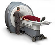
MRI tested in detection of iron stores in heart, liver and spleen, and pancreas
FRIDAY, Oct. 2, 2015 (HealthDay News) — Magnetic resonance imaging (MRI) is an accurate and safe tool for the detection of low levels of iron overload in patients with hereditary hemochromatosis, according to a letter to the editor published online Sept. 11 in the American Journal of Hematology.
Reijâne A. Assis, M.D., from Hospital Israelita Albert Einstein in São Paulo, Brazil, and colleagues examined all medical records of consecutive patients with iron overload seen in a tertiary hospital in Brazil (159 patients; 2008 to 2012) in order to correlate MRI T2* with HFE genetic profiles.
The researchers found that mutations in the HFE gene were identified in 68.6 percent of patients. Of the three patients (out of 126) with positive T2* MRIs, two had the H63D mutation (1 homozygous and 1 heterozygous). Of the 61 patients with liver overload, 27.9 percent were C282Y carriers and 50.8 percent carried the H63D mutation. The C282Y mutation (in either homozygosis or heterozygosis), compound heterozygous (C282/H63D), and H63D in homozygosis were significantly associated with a higher frequency of iron overload in the liver as measured by T2* (P = 0.001).
“Given the observation that MRI is an accurate and safe tool to measure iron stores in these organs, we believe that this technology should be incorporated in the investigation of suspected cases of hemochromatosis and contribute to guide therapeutic decisions such as phlebotomy,” the authors write.
Copyright © 2015 HealthDay. All rights reserved.


