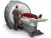
Functional magnetic resonance imaging with central sensitization beneficial for selecting analgesics
FRIDAY, Feb. 12, 2016 (HealthDay News) — Functional magnetic resonance imaging with central sensitization can be used in early human drug development, according to a study published in the January issue of Anesthesiology.
Vishvarani Wanigasekera, D.Phil., from the University of Oxford in the United Kingdom, and colleagues examined the utility of functional magnetic resonance imaging with capsaicin-induced central sensitization for differentiating an effective analgesic (gabapentin) from an ineffective analgesic and from placebo. Data were included for single oral doses in a double-blind phase I trial involving 24 participants.
The researchers found that suppression of the secondary mechanical hyperalgesia-evoked neural response in right nucleus cuneiformis and left posterior insular cortex and secondary somatosensory cortex was seen with gabapentin only. During central sensitization, suppression of the resting-state functional connectivity between the thalamus and secondary somatosensory cortex was also seen with gabapentin only, and was dependent on the plasma gabapentin level. A statistically significant difference between placebo and gabapentin was detected with a probability of ≥0.8 in a power analysis using neural activity from the left posterior insula and right nucleus cuneiformis. This was attenuated to ≤0.6 when using subjective pain ratings.
“Functional imaging with central sensitization can be used as a sensitive mechanism-based assay to guide go/no-go decisions on selecting analgesics effective in neuropathic pain in early human drug development,” the authors write.
Full Text (subscription or payment may be required)
Copyright © 2016 HealthDay. All rights reserved.


