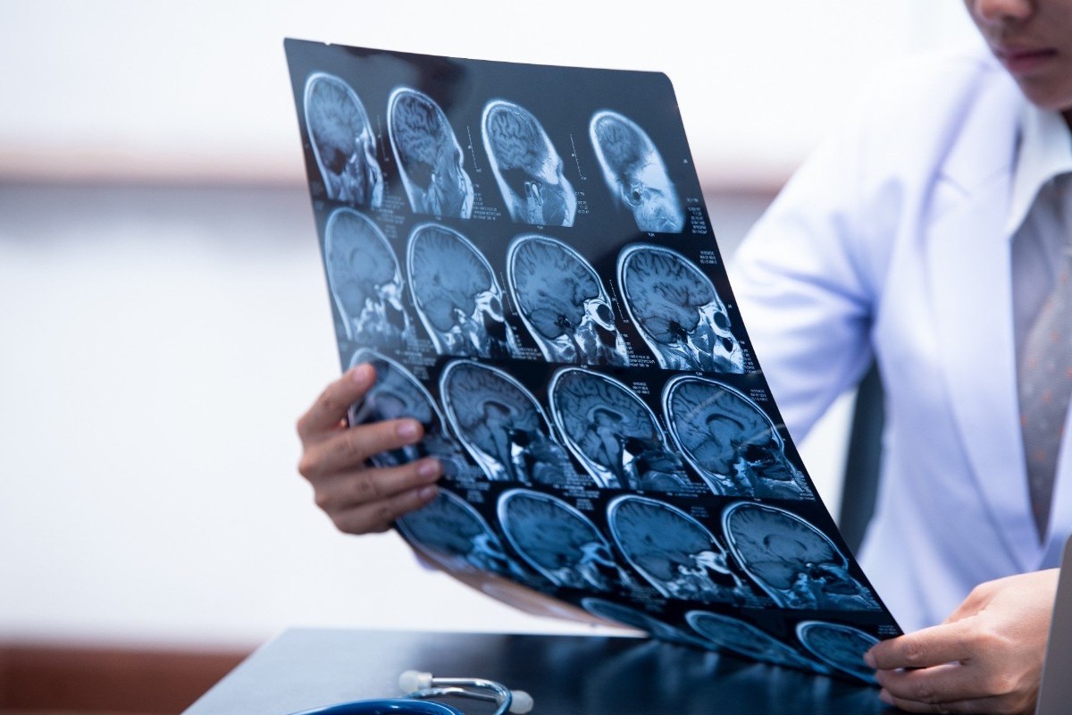
Existing clinical prognostic models significantly enhanced with DTI metrics
By Elana Gotkine HealthDay Reporter
FRIDAY, Aug. 16, 2024 (HealthDay News) — For patients with mild traumatic brain injury and normal computed tomography (CT), diffusion tensor imaging (DTI) improves existing prognostic models for functional outcome, according to a study published online Aug. 8 in eClinicalMedicine.
Sophie Richter, Ph.D., from the University of Cambridge in the United Kingdom, and colleagues examined whether existing prognostic models can be improved by serum biomarkers or DTI metrics from magnetic resonance imaging among 1,025 patients aged older than 18 years with a Glasgow Coma Score >12 and normal CT. The biomarkers glial fibrillary acidic protein (GFAP), neurofilament protein-light (NFL), and S100 calcium-binding protein B were obtained at a median of 8.8 hours, and DTI was performed at 13 days after injury.
The researchers found that 38 percent of the patients had incomplete recovery. The addition of biomarkers did not improve performance beyond the best existing clinical prognostic model (optimism-corrected area under the curve [AUC], 0.69; R2, 17 percent). All models were significantly enhanced with the addition of DTI metrics (best optimism-corrected AUC, 0.82; R2, 75 percent). Left posterior thalamic radiation, the left superior cerebellar peduncle, and right uncinate fasciculus were the top three prognostic tracts. In one of five DTI scans, serum biomarkers could have been avoided, with best performance for GFAP <12 hours and NFL at 12 to 24 hours.
“The problem is that the nature of concussion means patients and their general practitioners often don’t recognize that their symptoms are serious enough to need follow-up,” coauthor Virginia F. J. Newcombe, M.D., Ph.D., also from the University of Cambridge, said in a statement.
Several authors disclosed ties to the biopharmaceutical industry.
Copyright © 2024 HealthDay. All rights reserved.


