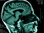
Alterations discern ADHD-I, ADHD-C, with discriminating features located in DMN, insular cortex
WEDNESDAY, Nov. 22, 2017 (HealthDay News) — Cerebral morphometric alterations can discriminate between children with attention-deficit/hyperactivity disorder (ADHD) and controls, according to a study published online Nov. 22 in Radiology.
Huaiqiang Sun, Ph.D., from the West China Hospital of Sichuan University in Chengdu, and colleagues performed anatomic and diffusion-tensor magnetic resonance imaging on a cohort of 83 age- and sex-matched children with newly diagnosed and never-treated ADHD and 87 healthy controls. For each participant, features representing the shape properties of gray matter and diffusion properties of white matter were extracted. To identify features with significant discriminative power for diagnosis and subtyping, the initial feature set was input into an all-relevant feature selection procedure within cross-validation loops.
The researchers observed no overall difference in terms of total brain volume or total gray and white matter volume for children with ADHD and control subjects. The mean classification accuracy achieved with classifiers was 73.7 percent for discrimination of patients with ADHD from controls. There was a significant contribution for alteration in cortical shape around the left temporal lobe, bilateral cuneus, and regions around the left central sulcus to group discrimination. The mean classification accuracy with classifiers was 80.1 percent for discriminating ADHD-inattentive subtype from ADHD-combined subtype (inattentive and hyperactive), with significant discriminating features located in the default mode network and insular cortex.
“By identifying features relevant for diagnosis and subtyping, these findings may advance the understanding of neurodevelopmental alterations related to ADHD,” the authors write.
One author disclosed financial ties to Takeda.
Copyright © 2017 HealthDay. All rights reserved.


