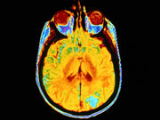
Imaging shows pattern of β-amyloid plaques that overlaps but differs from that of Alzheimer’s disease
THURSDAY, Feb. 4, 2016 (HealthDay News) — Imaging studies suggest that the development of β-amyloid (Aβ) plaques in individuals with traumatic brain injury (TBI) may be related to the presence of axonal damage, according to research published online Feb. 3 in Neurology.
Gregory Scott, M.B.B.S., of the Imperial College London, and colleagues assessed 28 individuals (nine with mild to moderate TBI recruited 11 months to 17 years after injury, nine controls, and 10 with Alzheimer’s disease) using 11C-Pittsburgh compound B (11C-PiB)-positron emission tomography and structural and diffusion magnetic resonance imaging studies. Binding potential (BPND) images of 11C-PiB were computed to index Aβ plaque density.
The researchers found that 11C-PiB BPND was increased in the posterior cingulate cortex and cerebellum in individuals with TBI compared with controls. Increased binding in the posterior cingulate cortex was observed with decreasing fractional anisotropy of associated white matter tracts and increased time since injury. Patterns of binding differed in those with TBI versus Alzheimer’s disease, with lower levels in neocortical regions and higher levels in the cerebellum.
“Increased Aβ burden was observed in TBI. The distribution overlaps with, but is distinct from, that of Alzheimer’s disease,” the authors write. “This suggests a mechanistic link between TBI and the development of neuropathologic features of dementia, which may relate to axonal damage produced by the injury.”
Several authors disclosed financial ties to the pharmaceutical and biomedical industries.
Copyright © 2016 HealthDay. All rights reserved.


