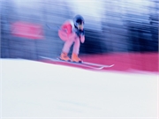
More distal femoral cortical irregularity found on knee MRI, mainly at medial head of gastrocnemius muscle
WEDNESDAY, June 17, 2020 (HealthDay News) — Young competitive alpine skiers have an increased prevalence of distal femoral cortical irregularities (DFCIs) on knee magnetic resonance imaging (MRI), according to a study published online June 16 in Radiology.
Christoph Stern, M.D., from Balgrist University Hospital in Switzerland, and colleagues compared the prevalence of DFCIs at different tendon attachment sites in youth competitive alpine skiers with the prevalence in young adults. From 2014 to 2019, unenhanced 3-T knee MRI scans obtained from 105 youth competitive alpine skiers were compared to images from 105 control participants of the same age by two radiologists.
The researchers identified DFCIs in 58 percent of skiers and 27 percent of control participants. Two of the skiers had more than one DFCI. For skiers and control participants, the distribution of DFCIs was at the medial head of the gastrocnemius muscle (MHG; 95.2 and 92.8 percent, respectively), at the lateral head of the gastrocnemius muscle (4.8 and 3.6 percent, respectively), and at the adductor magnus attachment site (0 and 3.6 percent, respectively). There was almost perfect interreader agreement (κ = 0.87). The mean size of MHG-related DFCIs did not differ for skiers versus control participants (3.7 versus 3.4 mm).
“DFCIs are benign lesions, and occurrence around the knee joint is associated with repetitive mechanical stress to the attachment sites of tendons into bone,” Stern said in a statement. “DFCIs should not be mistaken for malignancy and are not associated with intraarticular damage.”
Copyright © 2020 HealthDay. All rights reserved.


